Licensing

Attribution_nc_sa
Attribution Non-Commercial Share Alike:This image is licensed under a Creative Commons Attribution, Non-Commercial Share Alike License. View License Deed | View Legal Code
Comments: Powered by Pnyxe

Description

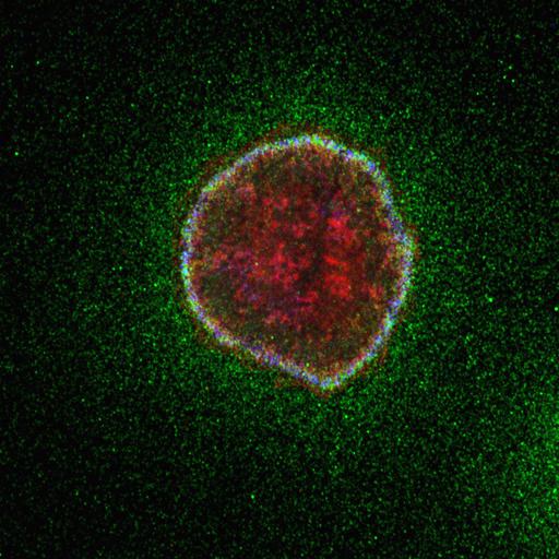
To determine the extent to which extracellular matrix proteins colocalize with the microtubule-anchoring factor LL5β, MCF-10A epithelial cells were immunostained with antibodies to the secreted autocrine laminin-5 (green), the laminin receptor integrin α6 (blue) and LL5β (red). Analyses suggest that LL5s are colocalized with activated pools of integrins associated with laminin-5. Overall findings from this publication showed that signaling from laminin-integrin associations plays a role in attaching microtubule plus ends to the epithelial basal cell cortex. This image is the original data file for Fig. 3 Panel D-b from J Cell Biol (2010) 189 (5):901-917.

Technical Details

MCF-10A epithelial cells were fixed with 1:1 mixture of methanol/acetone (-20° C, 10 min), permeabilized with 0.5% Triton in PBS, and immunostained sequentially with primary, then secondary antibodies. LL5β (Cy5, 633 nm laser), laminin-5 (rhodamine red-X, 543 nm laser)) and integrin α6 (Cy2, 488 nm laser, and observed using a confocal laser-scanning microscope system. For additional details see: J. Cell Biol. 2010. 189(5):901-917.

Biological Sources

NCBI Organism Classification
Homo sapiens
(human)
Cell Type
epithelial cell
Cell Line
MCF 10A
Cellular Component
microtubule
cell cortex part

Attribution

Names
Azusa Hotta
Tomomi Kawakatsu
Tomoya Nakatani
Toshitaka Sato
Chiyuki Matsui
Taiko Sukezane
Tsuyoshi Akagi
Tomoki Hamaji
Ilya Grigoriev
Anna Akhmanova
Yoshimi Takai
Yuko Mimori-Kiyosue
Published
J. Cell Biol. 2010. 189(5):901-917
Link
http://jcb-dataviewer.rupress.org/jcb/...
Pubmed
20513769

Grouping

This image is part of a group.

Imaging

Image Type
recorded image
Imaging Mode
confocal microscopy
Parameters Imaged
fluorescence emission
Source of Contrast
distribution of epitope
Visualization Methods
Cy2
Cy5
X-Rhodamine
Data Qualifiers
raw, unprocessed data
suitable for spatial measurements

Sample Preparation

Methods
methanol fixed tissue
permeabilized tissue
Relation To Intact Cell
dispersed cells in vitro

Dimensions

Spatial Axis Image Size Pixel Size
X 1024px 0.07μm
Y 1024px 0.07μm
*CIL – Cell Image Library accession number. Please use this to reference an image.