Alternate header for print version


Licensing
Public Domain: This image is in the public domain and thus free of any copyright restrictions. However, as is the norm in scientific publishing and as a matter of courtesy, any user should credit the content provider for any public or private use of this image whenever possible. Learn more
tweet  
*CIL – Cell Image Library accession number. Please use this to reference an image.

CIL:12068*  Cite 
Description

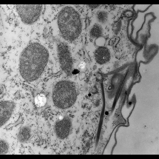
High resolution image. Basal bodies are supported by three fiber systems, a striated kinetodesmal fiber that arises from the proximal end of the basal body and curves upward and anteriorly within the longitudinal ridge to the cell’s right side of a row of basal bodies. Extending posteriorly and also curving up into the longitudinal ridge is a ribbon of postciliary microtubules. The kd may extend past several more anterior basal bodies but the postciliary ribbon does not extend beyond the next posterior basal body or pair of basal bodies in the kinety. Only the more posterior of the two basal bodies within a single depression is associated with the ends of these two accessory structures. An indentation of the plasma membrane, the parasomal sac, lies to the cell's right of each basal body or pair of basal bodies. TEM taken on 1/5/73 by R. Allen with Hitachi HU11A operating at 75kV. Neg. 19,500X. The raw film was scanned with an Epson Perfection V750 Pro. This image is best used for quantitative analysis. Standard glutaraldehyde fixation followed by osmium tetroxide, dehydrated in alcohol and embedded in an epoxy resin. Microtome sections prepared at approximately 75nm thickness. Additional information available at (http://www5.pbrc.hawaii.edu/allen/).

Biological Sources
NCBI Organism Classification
Paramecium multimicronucleatum
Cell Type
cell by organism
eukaryotic cell
Eukaryotic Protist
Ciliated Protist
Cellular Component
cell cortex
microtubule basal body
microtubule
coated pit
kinetodesmal fiber
Biological Context
Biological Process
cortical cytoskeleton organization
Attribution
Names
Richard Allen (University of Hawaii)
Link
http://www5.pbrc.hawaii.edu/allen/
Citation
Digital Object Identifier (DOI)
doi:10.7295/W9CIL12068
Archival Resource Key (ARK)
ark:/b7295/w9cil12068
Grouping This image is part of a group.
Imaging
Image Type
transmission electron microscopy (TEM)
illumination by electrons
Image Mode
detection of electrons
film
Parameters Imaged
electron density
Source of Contrast
stain with broad specificity
Visualization Methods
stain with broad specificity
osmium tetroxide
uranyl salt
lead salt
Processing History
recorded image
film
unprocessed raw data
Data Qualifiers
raw, unprocessed data
suitable for spatial measurements
Dimensions
Spatial Axis Image Size Pixel Size
X 4846px 0.77nm
Y 4590px 0.77nm