Alternate header for print version


Licensing
Attribution Non-Commercial; No Derivatives:This image is licensed under a Creative Commons Attribution, Non-Commercial, No Derivatives License. View License Deed | View Legal Code
tweet  
*CIL – Cell Image Library accession number. Please use this to reference an image.

CIL:11636*  Cite 
Description

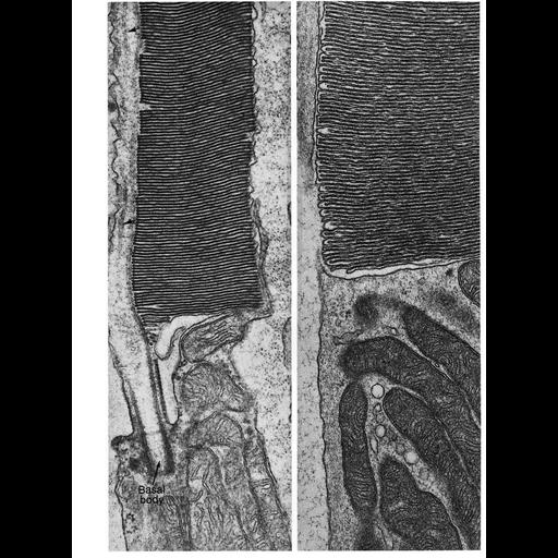
Figures 323 & 324 from Chapter 13 (Cilia and Flagella) of 'The Cell' by Don W. Fawcett M.D. The outer segments of the rods and cones of the vertebrate retina and many photoreceptors of invertebrates begin their differentiation as single cilia with a typical basal body and doublet microtubules forming a more or less complete axoneme. In their subsequent development, membranous discs containing the visual pigment accumulate within the confines of the ciliary membrane and become closely packed in parallel array to constitute the outer segment of the photoreceptor cell. Vestiges of the axonemal microtubules (at arrows) persist along one side of the outer segment which is connected to the inner segment by the proximal portion of the shaft of the original cilium. A copy of the chapter is available on the ASCB's BioEDUCATE website.

Biological Sources
NCBI Organism Classification
Rattus sp.
Cell Type
retinal cone cell
retinal rod cell
Cellular Component
cilium
mitochondrion
cytoplasm
Biological Context
Biological Process
epithelial cilium movement
cilium axoneme assembly
Attribution
Names
Don W. Fawcett
T. Kuwabara
Published
ISBN 0721635849
Link
http://www.ascb.org/bioeducate/FawcettTheCell...
Citation
Digital Object Identifier (DOI)
doi:10.7295/W9CIL11636
Archival Resource Key (ARK)
ark:/b7295/w9cil11636
Grouping This image is part of a group.
Imaging
Image Type
transmission electron microscopy (TEM)
illumination by electrons
Image Mode
detection of electrons
film
Parameters Imaged
electron density
Source of Contrast
stain with broad specificity
Visualization Methods
stain with broad specificity
osmium tetroxide
uranyl salt
lead salt
Processing History
recorded image
film
Print from negative scanned for Photoshop.
Data Qualifiers
processed data
Dimensions
Spatial Axis Image Size Pixel Size
X 960px ——
Y 1292px ——