Alternate header for print version


Licensing
Attribution Non-Commercial; No Derivatives:This image is licensed under a Creative Commons Attribution, Non-Commercial, No Derivatives License. View License Deed | View Legal Code
tweet  
*CIL – Cell Image Library accession number. Please use this to reference an image.

CIL:11634*  Cite 
Description

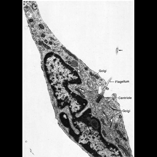
Figure 322 from Chapter 13 (Cilia and Flagella) of 'The Cell' by Don W. Fawcett M.D. Cilia and flagella usually occur on motile cells or on the free surfaces of epithelia. However, a solitary cilium is a normal occurrence on the cells lining the intra- hepatic bile ducts, the intercalated ducts of the pancreas, the rete testis, and certain segments of the nephron. The functional significance of these processes is not evident, since it seems unlikely that their motility could do more than create local turbulence in the fluid contents of these excretory ducts. Such abortive cilia usually lack the central pair of microtubules and are proba- bly immotile. Because modified cilia are found in photoreceptors and in olfactory epithelia, it has been suggested that these solitary cilia might have a sensory function but there is no evidence to support this speculation. It seems probable that they are merely anomalous, rudimentary structures with no function. A copy of the chapter is available on the ASCB's BioEDUCATE website.

Biological Sources
NCBI Organism Classification
Rattus sp.
Cell Type
mesenchymal cell
of the testis
Cellular Component
cilium
axoneme
nucleus
cytoplasm
Biological Context
Biological Process
stereocilium
microtubule basal body organization
Attribution
Names
Don W. Fawcett
Published
ISBN 0721635849
Link
http://www.ascb.org/bioeducate/FawcettTheCell...
Citation
Digital Object Identifier (DOI)
doi:10.7295/W9CIL11634
Archival Resource Key (ARK)
ark:/b7295/w9cil11634
Grouping This image is part of a group.
Imaging
Image Type
transmission electron microscopy (TEM)
illumination by electrons
Image Mode
detection of electrons
film
Parameters Imaged
electron density
Source of Contrast
stain with broad specificity
Visualization Methods
stain with broad specificity
osmium tetroxide
uranyl salt
lead salt
Processing History
recorded image
film
Print from negative scanned for Photoshop.
Data Qualifiers
processed data
Dimensions
Spatial Axis Image Size Pixel Size
X 898px ——
Y 1260px ——