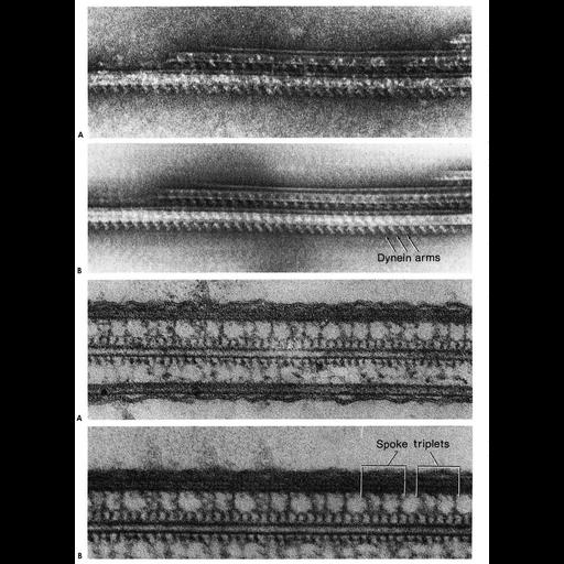
Figure 320 & 321 from Chapter 13 (Cilia and Flagella) of 'The Cell' by Don W. Fawcett M.D. The disposition of the dynein arms along the length of the doublets is studied to best advantage in dissociated cilia examined with negative staining. The upper figures show parts of three overlapping doublets from a demembranated cilium disintegrated in the presence of ATP. The second figure is a reinforced image of the same micrograph produced by a two-step linear translation. Free dynein arms can be seen projecting from the lowermost doublet at intervals of 24 nm along its length. Somewhat less clearly shown are arms bridging the interspaces between the three doublets. During active sliding, the space between doublets is considerably less than that usually seen in electron micrographs of thin cross sections. The microtubule sliding responsible for ciliary bending is believed to be due to attachment of arms on subunit A of one doublet to subunit B of the adjacent doublet followed by a conformational change resulting in arm deflection. The second set of figures are a thin longitudinal section of a cilium showing the radial spokes joining subunit A of a doublet to a row of projections along one of the central pair of microtubules. The spokes occur in groups of three with a repeat of 86 nm. The second figure is a reinforced image of the same micrograph resulting from linear translation in steps corresponding to 86nm between successive exposures. A copy of the chapter is available on the ASCB's BioEDUCATE website.
| Spatial Axis | Image Size | Pixel Size |
|---|---|---|
| X | 938px | —— |
| Y | 1276px | —— |