Alternate header for print version


Licensing
Attribution Non-Commercial; No Derivatives:This image is licensed under a Creative Commons Attribution, Non-Commercial, No Derivatives License. View License Deed | View Legal Code
tweet  
*CIL – Cell Image Library accession number. Please use this to reference an image.

CIL:11626*  Cite 
Description

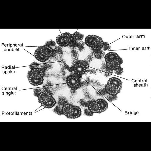
A figure from Chapter 13 (Cilia and Flagella) of 'The Cell' by Don W. Fawcett M.D. The microtubules of the axoneme are adequately preserved by routine fixation for electron microscopy of thin sections. These methods are less satisfactory for their subunits and appendages and have contributed little to our understanding of the organization of the ciliary matrix. Addition of tannic acid to the fixative results in an apparent thickening and a more intense staining of these components. The dynein arms are made more conspicuous, the radial spokes more robust, and the unstained protofilaments in the wall of the microtubules are clearly revealed in negative image. A copy of the chapter is available on the ASCB's BioEDUCATE website.

Biological Sources
NCBI Organism Classification
Rattus sp.
Cell Type
tracheal epithelial cell
Cellular Component
cilium
axoneme
microvillus
Biological Context
Biological Process
epithelial cilium movement
cilium axoneme assembly
Attribution
Names
Don W. Fawcett
Published
ISBN 0721635849
Link
http://www.ascb.org/bioeducate/FawcettTheCell...
Citation
Digital Object Identifier (DOI)
doi:10.7295/W9CIL11626
Archival Resource Key (ARK)
ark:/b7295/w9cil11626
Grouping This image is part of a group.
Imaging
Image Type
transmission electron microscopy (TEM)
illumination by electrons
Image Mode
detection of electrons
film
Parameters Imaged
electron density
Source of Contrast
stain with broad specificity
Visualization Methods
stain with broad specificity
osmium tetroxide
tannic acid
Processing History
recorded image
film
Scanned print from negative
Data Qualifiers
processed data
Dimensions
Spatial Axis Image Size Pixel Size
X 807px ——
Y 501px ——