Alternate header for print version


Licensing
Attribution Non-Commercial Share Alike:This image is licensed under a Creative Commons Attribution, Non-Commercial Share Alike License. View License Deed | View Legal Code
tweet  
*CIL – Cell Image Library accession number. Please use this to reference an image.

CIL:11623*  Cite 
Description

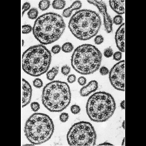
Abstracted from an image by Simionescu and Simionescu in The Journal of Cell Biology (70:608-621), ©1976, The Rockefeller University Press. This image was reprinted as Figure 319 from Chapter 13 (Cilia and Flagella) 'The Cell' by Don W. Fawcett M.D.(1981, W.B. Saunders Company). Ciliated cells of mammals often have microvilli interspersed among the cilia. These transverse sections near the cell surface permit a comparison of the size and internal structure of the two types of cell process. Both are limited by a trilaminar unit membrane and have a cytoplasmic matrix of similar density. The nonmotile microvilli have no resolvable internal structure, whereas the motile cilia have a consistent pattern of nine doublet microtubules uniformly spaced around a central pair of singlet microtubules. This nine plus two complex was visualized by the early light microscopists as a single axial fiber and was therefore called the axoneme. The term has been retained even though it does not accurately describe the microtubule complex revealed by the electron microscope. A copy of Chapter 13 from 'The Cell' is available on the ASCB's BioEDUCATE website.

Biological Sources
NCBI Organism Classification
Rattus sp.
Cell Type
tracheal epithelial cell
Cellular Component
cilium
axoneme
microvillus
Biological Context
Biological Process
epithelial cilium movement
cilium axoneme assembly
Attribution
Names
N. Simionescu
M. Simionescu
Published
J. Cell Biol. 70:608-621, 1976
Published
ISBN 0721635849
Link
http://jcb.rupress.org/content/70/3/622.abstr...
Fawcett's The Cell
Citation
Digital Object Identifier (DOI)
doi:10.7295/W9CIL11623
Archival Resource Key (ARK)
ark:/b7295/w9cil11623
Grouping This image is part of a group.
Imaging
Image Type
transmission electron microscopy (TEM)
illumination by electrons
Image Mode
detection of electrons
film
Parameters Imaged
electron density
Source of Contrast
stain with broad specificity
Visualization Methods
stain with broad specificity
osmium tetroxide
tannic acid
Processing History
recorded image
film
Print from negative scanned for Photoshop.
Data Qualifiers
processed data
Dimensions
Spatial Axis Image Size Pixel Size
X 898px ——
Y 1256px ——