Alternate header for print version


Licensing
Attribution Non-Commercial; No Derivatives:This image is licensed under a Creative Commons Attribution, Non-Commercial, No Derivatives License. View License Deed | View Legal Code
tweet  
*CIL – Cell Image Library accession number. Please use this to reference an image.

CIL:11491*  Cite 
Description

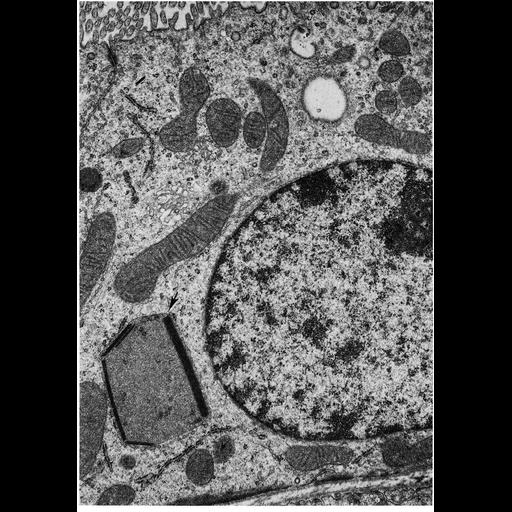
Figure 288 from Chapter 9 (Peroxisomes) of 'The Cell, 2nd Ed.' by Don W. Fawcett M.D. Micrograph of a cell from the renal proximal convoluted tubule of the rat. A peroxisome (arrow) near the base of the cell contains both crystals and a few circular profiles of cylinders in cross section. Image courtesy of Michael Barrett and Paul Heidger (1975) Cell and Tissue 157:283-305, PMID: 1122543. A PDF copy of the accompanying chapter is available on the ASCB’s BioEDUCATE website.

Biological Sources
NCBI Organism Classification
Rattus
Cell Type
renal principal cell
Cellular Component
peroxisome
cytoplasm
organelle
Biological Context
Biological Process
cellular catabolic process
Attribution
Names
Don W. Fawcett
Michael Barrett
Paul Heidger
Published
ISBN 0721635849
Link
http://www.ascb.org/bioeducate/FawcettTheCell...
Citation
Digital Object Identifier (DOI)
doi:10.7295/W9CIL11491
Archival Resource Key (ARK)
ark:/b7295/w9cil11491
Grouping This image is part of a group.
Imaging
Image Type
transmission electron microscopy (TEM)
Image Mode
illumination by electrons
detection of electrons
Parameters Imaged
electron density
Source of Contrast
stain with broad specificity
Data Qualifiers
processed data
Sample Preparation
Methods
crosslinking-fixative fixed tissue
embedded tissue
Relation To Intact Cell
microtome-sectioned tissue
Dimensions
Spatial Axis Image Size Pixel Size
X 894px ——
Y 1280px ——