Alternate header for print version


Licensing
Attribution Non-Commercial; No Derivatives:This image is licensed under a Creative Commons Attribution, Non-Commercial, No Derivatives License. View License Deed | View Legal Code
tweet  
*CIL – Cell Image Library accession number. Please use this to reference an image.

CIL:11467*  Cite 
Description

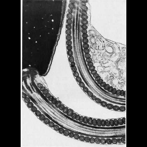
The midpiece of spermatzoa in nearly all vertebrates is enclosed in a sheath of mitochondria, which provide a ready source of ATP necessary for sperm motility. Here, a longitudinal section through spermatozoa of the dormouse, Glis glis, shows elongate mitochondria arranged end to end in a helix, an organization common in mammals. This image by David Phillips is Figure 261 from Chapter 7 (Mitochondria) of 'The Cell, 2nd Ed.' by Don W. Fawcett M.D. A PDF copy of the accompanying chapter is available on the ASCB’s BioEDUCATE website.

Biological Context
Biological Process
sperm motility
oxidative phosphorylation
ATP biosynthetic process
Attribution
Names
David Phillips
Published
ISBN 0721635849
Link
Fawcett's "The Cell" 2nd Ed.
Citation
Digital Object Identifier (DOI)
doi:10.7295/W9CIL11467
Archival Resource Key (ARK)
ark:/b7295/w9cil11467
Grouping This image is part of a group.
Imaging
Image Type
transmission electron microscopy (TEM)
Image Mode
illumination by electrons
detection of electrons
Parameters Imaged
electron density
Source of Contrast
stain with broad specificity
Data Qualifiers
processed data
Sample Preparation
Methods
crosslinking-fixative fixed tissue
embedded tissue
Relation To Intact Cell
microtome-sectioned tissue
Dimensions
Spatial Axis Image Size Pixel Size
X 891px ——
Y 1240px ——