Alternate header for print version


Licensing
Attribution Non-Commercial; No Derivatives:This image is licensed under a Creative Commons Attribution, Non-Commercial, No Derivatives License. View License Deed | View Legal Code
tweet  
*CIL – Cell Image Library accession number. Please use this to reference an image.

CIL:11099*  Cite 
Description

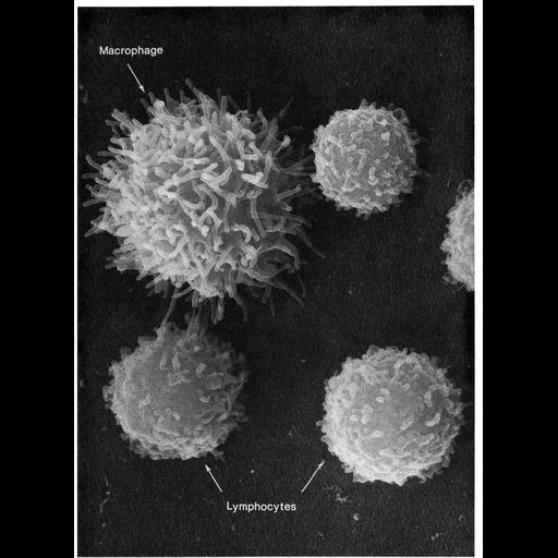
Cells from peritoneal cavity exudate of the mouse, exposed to sodium azide at 22°C for 22 minutes. The macrophage in the field of view has extended long microvilli, showing a rapid response to changes in metabolic and environmental conditions. Image by Emma Shelton, Figure 35 from Chapter 2 (Specializations of the Free Surface) of 'The Cell, 2nd Ed.' by Don W. Fawcett M.D. A PDF copy of the accompanying chapter is available on the ASCB’s BioEDUCATE website.

Biological Sources
NCBI Organism Classification
Mus musculus
Cell Type
macrophage
lymphocyte
Cellular Component
microvillus
Biological Context
Biological Process
immune system process
Attribution
Names
Emma Shelton
Published
ISBN 0721635849
Link
Fawcett's "The Cell, 2nd Ed."
Citation
Digital Object Identifier (DOI)
doi:10.7295/W9CIL11099
Archival Resource Key (ARK)
ark:/b7295/w9cil11099
Grouping This image is part of a group.
Imaging
Image Type
scanning electron microscopy (SEM)
Image Mode
illumination by electrons
secondary_electron imaging
Parameters Imaged
secondary electron generation
Data Qualifiers
processed data
Sample Preparation
Methods
critical_point dried specimen
Relation To Intact Cell
dispersed cells in vitro
Dimensions
Spatial Axis Image Size Pixel Size
X 918px ——
Y 1252px ——