Alternate header for print version


Licensing
Attribution Non-Commercial; No Derivatives:This image is licensed under a Creative Commons Attribution, Non-Commercial, No Derivatives License. View License Deed | View Legal Code
tweet  
*CIL – Cell Image Library accession number. Please use this to reference an image.

CIL:11052*  Cite 
Description

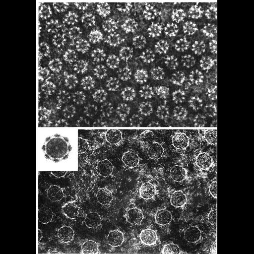
Early transmission electron micrographs showing pore details in negatively stained preparations of nuclear envelopes. Upper panel shows the pronounced 8-fold symmetry in the closely spaced pores of the oocyte of the newt Taricha granulosa. Lower panel depicts the oocyte envelope of the newt Notophthalmus viridescens revealing 8-fold symmetry upon rotational averaging (inset). (Upper micrograph from A. Faberge (1973) Z. zellforsch. 136: 183-190, reprinted with permission as Fig. 151; lower micrograph from Gall (1967) J. Cell Biol 32: 391, reprinted with permission as Fig. 152 from Chapter 4 (Nucleus) of 'The Cell, 2nd Ed.' by Don W. Fawcett M.D. A PDF copy of the accompanying chapter is available on the ASCB's BioEDUCATE website.

Technical Details

See: AC Faberge 1973 Direct demonstration of 8-fold symmetry in nuclear pores. Z Zellforsch Mikrosk Anat 136:183-190. J. Gall 1967 Octagonal nuclear pores J Cell Biol 32:391-399

Biological Sources
NCBI Organism Classification
Taricha granulosa
Notophthalmus viridescens
Cell Type
oocyte
Cellular Component
nuclear envelope
nuclear pore
Biological Context
Biological Process
nucleus organization
nuclear transport
nuclear pore organization
Attribution
Names
A.C. Faberge
Joseph Gall
Published
AC Faberge 1973 Direct demonstration of 8-fold symmetry in nuclear pores. Z Zellforsch Mikrosk Anat 136:183-190
Published
J. Gall 1967 Octagonal nuclear pores J Cell Biol 32:391-399
Published
from: Don W. Fawcett M.D ‘The Cell’ 2nd edition, W.B. Saunders Chapter 4 (Nucleus)
Published
ISBN 0721635849
Pubmed
4119506
Pubmed
10976230
Link
Pubmed
Journal of Cell Biology
ASCB Bioeducate Program
Citation
Digital Object Identifier (DOI)
doi:10.7295/W9CIL11052
Archival Resource Key (ARK)
ark:/b7295/w9cil11052
Grouping This image is part of a group.
Imaging
Image Type
film
Image Mode
transmission electron microscopy (TEM)
detection of electrons
Parameters Imaged
electron density
Source of Contrast
differences in adsorption or binding of stain
Visualization Methods
negative staining
tungsten compound
Processing History
Image extracted from .pdf file of 'The Cell'
Data Qualifiers
processed data
Sample Preparation
Methods
unembedded tissue
Relation To Intact Cell
isolated subcellular component
Dimensions
Spatial Axis Image Size Pixel Size
X 908px ——
Y 1272px ——