Alternate header for print version


Licensing
Attribution Non-Commercial; No Derivatives:This image is licensed under a Creative Commons Attribution, Non-Commercial, No Derivatives License. View License Deed | View Legal Code
tweet  
*CIL – Cell Image Library accession number. Please use this to reference an image.

CIL:11023*  Cite 
Description

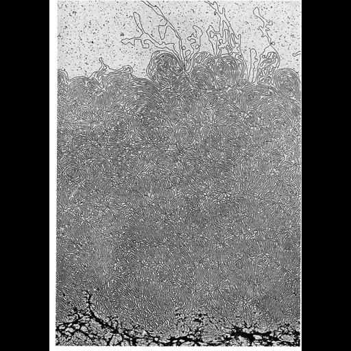
Transmission electron micrograph showing part of a decondensed mitotic chromosome. The residual protein 'scaffold' (at bottom) is surrounded by a 'halo' of DNA loops 20 to 24 micrometers long. The entire chromosome is seen in the grouped CIL image 11021.

Technical Details

Micrograph from J.R. Paulson and U.K. Laemmli (1977) Cell, 12:817-828, and republished with permission as Figure 131 from Chapter 4 (Nucleus) of 'The Cell' by Don W. Fawcett M.D. This image is the enlarged inset panel from CIL:11021 in this image group.

Biological Sources
NCBI Organism Classification
Homo sapiens
Cell Line
HeLa
Cellular Component
nuclear chromosome
chromatin
Attribution
Names
J. R. Paulson
U. K. Laemmli
Don W Fawcett
Published
ISBN 0721635849
Published
from: Don W. Fawcett M.D ‘The Cell’ 2nd edition, W.B. Saunders Chapter 4 (Nucleus)
Pubmed
922894
Link
ASCB BioEDUCATE Program
Cell
Citation
Digital Object Identifier (DOI)
doi:10.7295/W9CIL11023
Archival Resource Key (ARK)
ark:/b7295/w9cil11023
Grouping This image is part of a group.
Imaging
Image Type
film
Image Mode
transmission electron microscopy (TEM)
detection of electrons
Parameters Imaged
electron density
Source of Contrast
differences in adsorption or binding of stain
Visualization Methods
uranyl salt
Processing History
Image extracted from .pdf file of 'The Cell'
Data Qualifiers
processed data
Sample Preparation
Methods
unfixed tissue
Relation To Intact Cell
isolated subcellular component
Dimensions
Spatial Axis Image Size Pixel Size
X 918px ——
Y 1276px ——