Alternate header for print version


Licensing
Public Domain: This image is in the public domain and thus free of any copyright restrictions. However, as is the norm in scientific publishing and as a matter of courtesy, any user should credit the content provider for any public or private use of this image whenever possible. Learn more
tweet  
*CIL – Cell Image Library accession number. Please use this to reference an image.

CIL:10455*  Cite 
Description

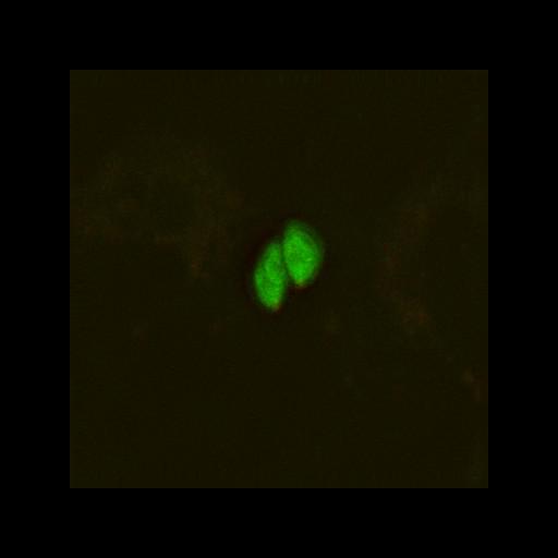
Toxoplasma gondii double stable clone expressing TgAPR1-mCherryFP (an MTOC protein) and EGFP-beta1-tubulin. 3D stack image of a live unfixed specimen acquired using API Delta Vision on Olympus ix70 wide-field microscope with Cool SNAP HQ/ICX285 CCD camera. Filter set: EGFP excitation 450-490 and emission 500-540; mCherry excitation 555-592 and emission: 600-665. Dichromatic mirror: Chroma 89021. Objective: Olympus 60x water immersion, 1.2 NA. Deconvolved stack corresponds to raw data: CIL# 10454 and raw data with DIC: CIL# 10460. Unpublished image that is similar to images published in PMID: 16518471. See publication for other experimental details.

Biological Sources
NCBI Organism Classification
Toxoplasma gondii RH
Cell Type
Toxoplasma gondii RH
Cell Line
Toxoplasma gondii RH double stable clone expressing TgAPR1-mCherryFP and EGFP-beta1-tubulin
Cellular Component
cortical microtubule, transverse to long axis
microtubule organizing center
Biological Context
Biological Process
regulation of cell shape
Molecular Function
structural constituent of cytoskeleton
Human Disease
toxoplasmosis
Attribution
Names
John Murray
Citation
Digital Object Identifier (DOI)
doi:10.7295/W9CIL10455
Archival Resource Key (ARK)
ark:/b7295/w9cil10455
Grouping This image is part of a group.
Imaging
Image Type
recorded image
charge coupled device (CCD)
Image Mode
fluorescence microscopy
Parameters Imaged
fluorescence emission
Source of Contrast
distribution of a specific protein
Visualization Methods
EGFP
mCherryFP
Processing History
3D-deconvolution
Data Qualifiers
processed data
suitable for spatial measurements
Sample Preparation
Methods
living tissue
Relation To Intact Cell
whole mounted tissue
Dimensions
Spatial Axis Image Size Pixel Size
X 384px 106.7nm
Y 384px 106.7nm
Z 15px 300nm