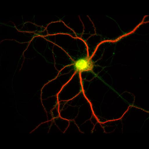
Cultured hippocampal neurons after 23 days in vitro, immunostained for MAP2, a microtubule associated protein localized to dendrites (red), and PSD95, a scaffolding protein concentrated in the postsynaptic density of excitatory synapses, particularly in dendritic spines (green). Detailed methods: Embryonic rat hippocampal neurons were prepared as previously described (see Kaech and Banker, 2006, Nat Protoc). Cells were prepared for fluorescent staining as previously described (Withers and Banker, 1998, in Culturing Nerve Cells, MIT Press). Briefly, cells were fixed (4% formaldehyde, 4% sucrose in phosphate buffered saline, pH 7.4), permeabilized with 0.25% Triton and immunostained for MAP2 (from S. Halpain) with d549 conjugated secondary, excitation, 555, emission, 568, Jackson Immunoresearch) and PSD95 (from NeuroMab, with Alexa488 conjugated secondary, excitation, 496, emission, 519, Invitrogen). Images were acquired with a Leica DMRA microscope with a 40X lens (HCX PL Fluotar, NA 0.75), Photometrics CoolSnap ES CCD camera and MetaMorph software. The merged image was generated by creating a MetaMorph stack file.
| Spatial Axis | Image Size | Pixel Size |
|---|---|---|
| X | 1300px | 0.167µm |
| Y | 1030px | 0.167µm |