Alternate header for print version


Licensing
Public Domain: This image is in the public domain and thus free of any copyright restrictions. However, as is the norm in scientific publishing and as a matter of courtesy, any user should credit the content provider for any public or private use of this image whenever possible. Learn more
tweet  
*CIL – Cell Image Library accession number. Please use this to reference an image.

CIL:10103*  Cite 
Description

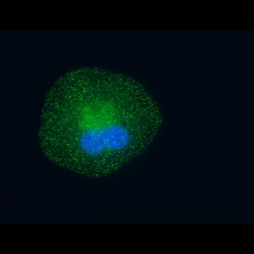
Cultured retinal pigment epithelial cells immunofluorescently labeled for adaptor protein-2 (AP2) (green) and nucleus (blue). The cells were fixed in 2% PFA and 0.5% Triton X-100 for 2 minutes followed by post-fixation 4% PFA. AP2 was detected with AP-6 primary antibody and secondary FITC antibody. The nucleus was detected with DAPI staining. Images were collected on an Olympus IX-71 epifluorescence microscope using a 100X 1.4 NA objective with 4.500ms exposure for AP2 and 50ms exposure for DAPI (67nm/pixel).

Biological Sources
NCBI Organism Classification
Homo sapiens
Cell Type
epithelial cell
Cell Line
ARPE-19
Cellular Component
AP-2 adaptor complex
nucleus
Attribution
Names
Allen Liu
Sandra L. Schmid
Citation
Digital Object Identifier (DOI)
doi:10.7295/W9CIL10103
Archival Resource Key (ARK)
ark:/b7295/w9cil10103
Sample Preparation
Methods
formaldehyde fixed tissue
permeabilized tissue
Relation To Intact Cell
dispersed cells in vitro
Dimensions
Spatial Axis Image Size Pixel Size
X 1344px 67nm
Y 1024px 67nm
Channel Wavelength
1 350, 488nm