Alternate header for print version


Licensing
Public Domain: This image is in the public domain and thus free of any copyright restrictions. However, as is the norm in scientific publishing and as a matter of courtesy, any user should credit the content provider for any public or private use of this image whenever possible. Learn more
tweet  
*CIL – Cell Image Library accession number. Please use this to reference an image.

CIL:10061*  Cite 
Description

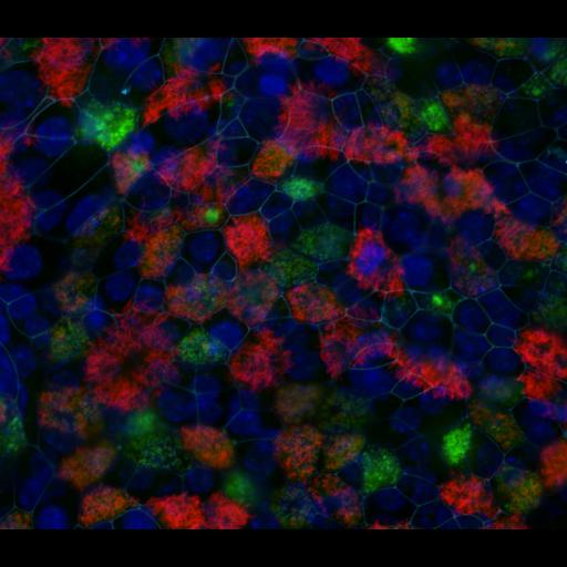
Mouse tracheal epithelia cells (MTEC) were grown at air-liquid interface to induce differentiation, centriole amplification and ciliogenesis. Cells were fixed with ice-cold methanol for 10 min. Samples were stained with antibodies against acetylated tubulin to mark cilia (red), the centriolar protein Cep120 to mark basal bodies (green), and anti-ZO-1 to mark the cell-cell junctions (light blue). Secondary antibodies were Alexa 594 for acetylated tubulin, Alexa 488 for Cep120, and Cy5 for Zo-1. Nuclei were stained using DAPI (blue). Images were captured using Openlab 4.0.4 software controlling an Axiovert 200M microscope (Carl Zeiss, Inc.) with a 100X 1.4 NA objective.

Biological Sources
NCBI Organism Classification
Mus musculus
Cell Type
tracheal epithelial cell
Cellular Component
cell-cell junction
microtubule basal body
nucleus
acetylated tubulin
Biological Context
Biological Process
lung ciliated cell differentiation
Attribution
Names
Moe Mahjoub
Tim Stearns
Citation
Digital Object Identifier (DOI)
doi:10.7295/W9CIL10061
Archival Resource Key (ARK)
ark:/b7295/w9cil10061
Grouping This image is part of a group.
Sample Preparation
Methods
methanol fixed tissue
Relation To Intact Cell
detergent permeabilized
Dimensions
Spatial Axis Image Size Pixel Size
X 1171px 0.094µm
Y 1017px 0.094µm
Channel Wavelength
1 350, 488, 594, 633nm