Alternate header for print version


Licensing
Public Domain: This image is in the public domain and thus free of any copyright restrictions. However, as is the norm in scientific publishing and as a matter of courtesy, any user should credit the content provider for any public or private use of this image whenever possible. Learn more
tweet  
*CIL – Cell Image Library accession number. Please use this to reference an image.

CIL:10007*  Cite 
Description

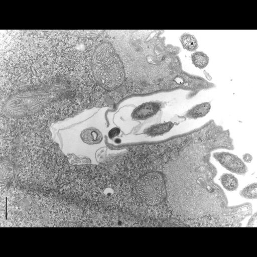
An image of one of several contractile vacuole pores of the proter of a dividing cell. This is presumably at an early stage of development as division is not yet complete. This section shows the pore to be encased in microtubules. The micrograph shows the microtubules cut transversely on one side so one can count them and obliquely on the other side of the pore. Whether the CV is yet functional at this stage is not known although the pore septum appears as two flaps of membrane. The pore lies close to the division furrow in the proter. TEM taken on 5/21/69 by M. Sage with Philips 300 operating at 60kV. Neg. 14,800X.Bar = 0.5µm. The negative was printed to paper and the image was scanned to Photoshop. This digitized image is available for qualitative analysis. A raw, unprocessed, high resolution version of this image (CIL:4666) is in the library and available for quantitative analysis. Standard glutaraldehyde fixation followed by osmium tetroxide, dehydrated in alcohol and embedded in an epoxy resin. Microtome sections prepared at approximately 75nm thickness. Additional information available at (http://www5.pbrc.hawaii.edu/allen/).

Biological Sources
NCBI Organism Classification
Didinium nasutum
Cell Type
eukaryotic cell
Eukaryotic Protist
Ciliated Protist
Cell Line
Carolina Biological Supply Company, NC, U.S.A.
Cellular Component
contractile vacuole pore
cell cortex
cell cortex part
Biological Context
Biological Process
water transport
contractile vacuole development
Attribution
Names
M. Sage
Richard Allen
Link
http://www5.pbrc.hawaii.edu/allen/
Citation
Digital Object Identifier (DOI)
doi:10.7295/W9CIL10007
Archival Resource Key (ARK)
ark:/b7295/w9cil10007
Grouping This image is part of a group.
Imaging
Image Type
transmission electron microscopy (TEM)
illumination by electrons
Image Mode
detection of electrons
film
Parameters Imaged
electron density
Source of Contrast
stain with broad specificity
Visualization Methods
stain with broad specificity
osmium tetroxide
uranyl salt
lead salt
Processing History
film
Print from negative scanned for Photoshop.
Data Qualifiers
processed data
suitable for spatial measurements
Dimensions
Spatial Axis Image Size Pixel Size
X 3448px ——
Y 2656px ——