Alternate header for print version
Advanced search
Contributors
Help
Submit
Search
menu
Cell Process
Cell Component
Cell Type
Organism
Microbial
Alzheimer's
Data Sets
University of California, San Diego
9500 Gilman Drive
La Jolla, CA 92093-0608, USA
Voice
: (858) 534-0276
Fax
: (858) 534-7497
Email
: dorloff@ncmir.ucsd.edu
Grouped images - the images shown below are related
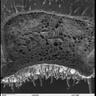
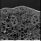
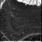
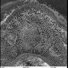
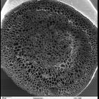
CIL:40378
NCBI Organism Classification
Solenostemon scutellarioides
Biological Process
plant stem organization
Cellular Component
cortex
Scanning electron microscope image of cross-section through a Solenostemon scute...
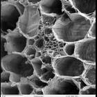
CIL:40379
NCBI Organism Classification
Solenostemon scutellarioides
Biological Process
vascular bundle organization
Cellular Component
none specified
Scanning electron microscope image of cross-section through a Solenostemon scute...
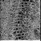
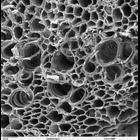
CIL:40380
NCBI Organism Classification
Solenostemon scutellarioides
Biological Process
plant stem organization
Cellular Component
cortex
Scanning electron microscope image of cross-section through a Solenostemon scute...
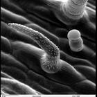
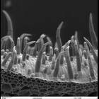
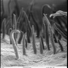
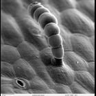
CIL:40381
NCBI Organism Classification
Solenostemon scutellarioides
Biological Process
epidermis morphogenesis
Cellular Component
trichome
Scanning electron microscope image of the epidermal surface of a Solenostemon sc...
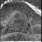
CIL:40382
NCBI Organism Classification
Solenostemon scutellarioides
Biological Process
epidermis morphogenesis
Cellular Component
trichome
Scanning electron microscope image of cross-section through a Solenostemon scute...
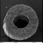
CIL:40383
NCBI Organism Classification
Oryza sativa
Biological Process
plant stem organization
Cellular Component
none specified
Scanning electron microscope image of a cross-section through an Oryza sativa (R...
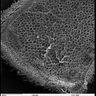
CIL:40384
NCBI Organism Classification
Oryza sativa
Biological Process
plant stem organization
Cellular Component
cell cortex
Scanning electron microscope image of Oryza sativa (Rice) stem. This is a high ...
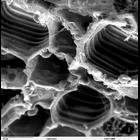
CIL:40385
NCBI Organism Classification
Oryza sativa
Biological Process
plant stem organization
Cellular Component
starch grain
Scanning electron microscope image of longitudinal section of Oryza sativa (rice...
CIL:40386
NCBI Organism Classification
Psidium guajava
Biological Process
plant stem organization
Cellular Component
mucilage
Scanning electron microscope image of cross-section through a Psidium guajava st...
CIL:40387
NCBI Organism Classification
Psidium guajava
Biological Process
plant stem organization
Cellular Component
none specified
High magnification scanning electron micrograph of a cross-section through a Psi...
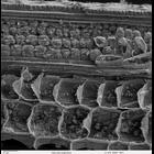
CIL:40388
NCBI Organism Classification
Psidium guajava
Biological Process
epidermis morphogenesis
Cellular Component
trichome
Scanning electron microscope image of Psidium guajava epidermal surface of the s...
CIL:40389
NCBI Organism Classification
Helianthus annuus
Biological Process
plant stem organization
Cellular Component
cortex
Scanning electron microscope image of cross-section through Helianthus annuus (s...
CIL:40390
NCBI Organism Classification
Helianthus annuus
Biological Process
plant stem organization
Cellular Component
cortex
Scanning electron microscope image of cross-section through Helianthus annuus (s...
CIL:40391
NCBI Organism Classification
Helianthus annuus
Biological Process
plant stem organization
Cellular Component
none specified
Scanning electron microscope image of a cross-section through a Helianthus annuu...
CIL:40392
NCBI Organism Classification
Helianthus annuus
Biological Process
epidermis morphogenesis
Cellular Component
trichome
Scanning electron microscope image of Helianthus annuus (sunflower) stem epiderm...
CIL:40393
NCBI Organism Classification
Helianthus annuus
Biological Process
plant stem organization
Cellular Component
none specified
Scanning electron microscope image of a longitudinal section of a Helianthus ann...
CIL:40394
NCBI Organism Classification
Tradescantia virginiana
Biological Process
plant stem organization
Cellular Component
none specified
Scanning electron microscope image of a cross-section through a Tradescantia vir...
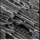
CIL:40395
NCBI Organism Classification
Tradescantia virginiana
Biological Process
plant stem organization
Cellular Component
none specified
Scanning electron microscope image of showing the phloem and xylem in a vascular...